The message was never missing. The channel was.
Apraxia is the gap between what a child intends and what the body delivers. In autism that gap is measurable, it is larger than basic coordination explains, and it is routinely mistaken for absence of thought. This is what is established, what is contested, and what nobody has measured yet.
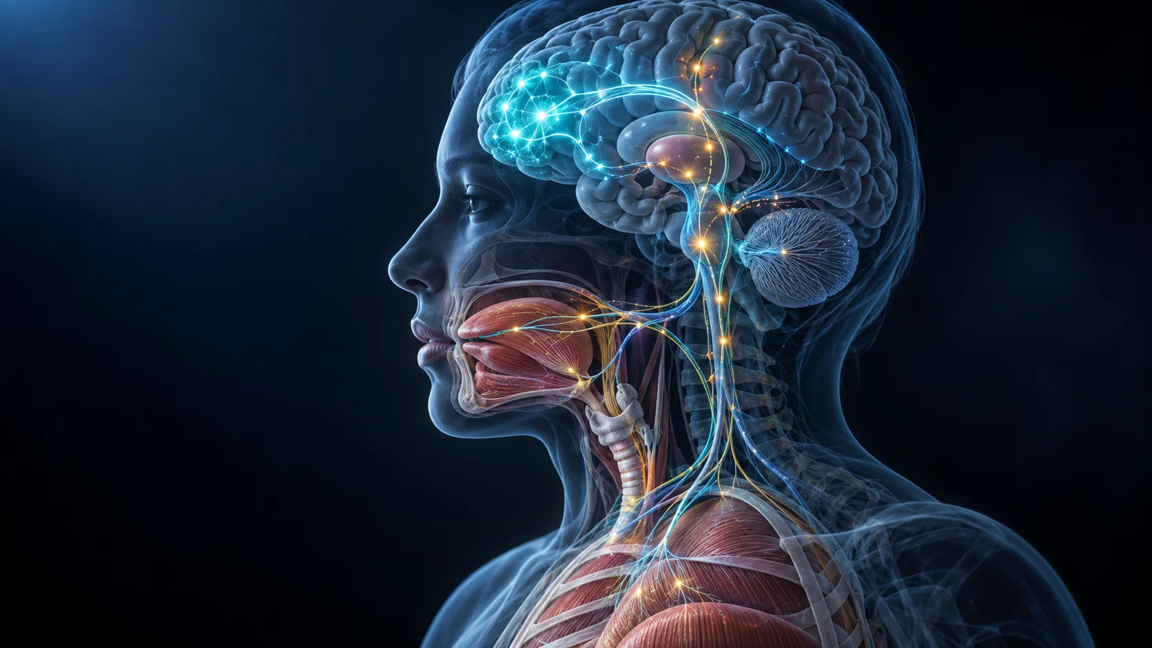
A child who cannot show you what they know.
Praxis is the ability to plan a movement and then release it on demand. It is not strength, and it is not coordination. It is the step between having the plan and issuing it.
Ask a child to wave goodbye and nothing happens. 10 minutes later, unprompted, the same child waves perfectly at a car pulling out of the driveway. The movement exists. Access to it, on request, in that moment, does not.
Parents describe this constantly, and it is one of the most misread things in autism. A child who cannot answer a question on cue is recorded as not understanding the question. A child whose mouth will not produce the word is recorded as not having the word.
This is measurable. In autistic children, performance on praxis testing is impaired well beyond what basic motor skill accounts for, and the size of that impairment tracks autism symptom severity (Dziuk, 2007). It is not explained by attention problems either: children with ADHD do not show the same pattern (MacNeil and Mostofsky, 2012).
A meta-analysis of motor coordination in autism found a standardized mean difference of 1.20, which is a large effect, not a footnote to the diagnosis (Fournier, 2010).
Read this caveat before the rest of the page
Nearly all of the praxis research above was done in speaking, school age children with average IQ. Almost none of it tested nonspeaking children, who are the children this question matters to most. That gap runs through this entire literature, and it is one of the strongest reasons to keep reading this page as a hypothesis rather than a conclusion.
What DMT actually is.
N,N-dimethyltryptamine is serotonin's structural cousin with 2 methyl groups added to the nitrogen at the end of its side chain. That small change is what makes it a potent agonist at the serotonin 2A receptor.
Start with tryptophan, an amino acid from food. Remove its carboxyl group and you get tryptamine. Add 2 methyl groups to tryptamine and you get DMT. Both steps are ordinary biochemistry, and both happen in living systems.
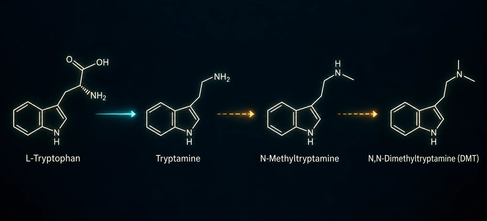
DMT has been detected in mammalian tissue since the 1960s. The important question has never been whether the body can make it. The question is how much, where, and whether the amount is enough to change how a brain works.
Here the honest answer is uncomfortable. The best direct measurement in a living brain found extracellular DMT in rat visual cortex at roughly 0.05 to 2.2 nM, which is the same order of magnitude as serotonin measured in the same animals at 2.10 nM (Dean, 2019).
But the pharmacology community reads that as too little to matter: trace amounts are present, and they are "not sufficient to produce psychoactive effects" (Nichols, 2017). Producing a psychedelic effect with DMT from outside the body takes plasma levels of roughly 12 to 90 ng/mL, and no one has ever quantified endogenous DMT in human brain tissue (Barker, 2018).
What this means for everything that follows
Presence is established. Sufficiency is not. Any model that depends on endogenous DMT doing real work in a human brain is, at this moment, unproven at its most basic quantitative step. We state that at the top rather than at the bottom.
Bacteria that turn tryptophan into tryptamine.
Some gut organisms carry a tryptophan decarboxylase, the enzyme that performs the first step. It is a genuinely unusual trait.
The defining work identified 2 bacterial tryptophan decarboxylases, in Ruminococcus gnavus and Clostridium sporogenes, described the activity as exceedingly rare among bacteria, and estimated that at least 10% of people carry an organism with one (Williams, 2014). Later screening added Blautia hansenii, Enterocloster asparagiformis and Clostridium nexile, finding tryptamine production in 5 of 13 strains tested (Otaru, 2024).
That number, 5 of 13, is the part most often lost in translation. This is a strain level trait, not a genus level one. Most close relatives of these organisms produce nothing at all (Gonçalves, 2022). A stool report showing a genus is elevated does not establish that the enzyme is present, and we should stop writing as though it does.
205 samples
Sequencing data from 205 stool samples across 172 people in this practice. These are the verified figures, and they replace the larger numbers quoted in earlier drafts of this argument.
16 / 50 / 66%
R. gnavus elevated in 16%. Blautia at genus level in 50%. Clostridium at genus level in 66%. Genus level readings, with the caveat above.
83%
83% of children showed at least one of the 3 elevated. Striking, and still not proof of decarboxylase capacity, which is the measurement that would settle it.
The prediction this makes
If the pathway is driven by the enzyme rather than by any single organism, then tryptamine output should track total decarboxylase capacity across all taxa present, not R. gnavus abundance on its own. That is a falsifiable claim, and it can be tested against stool data that already exists.
Getting out of the gut. The contested step.
For a bacterial metabolite to matter systemically it has to leave the intestine. The idea that intestinal permeability is increased in autism has real support, and real opposition, and anyone presenting only one side is selling something.
For: raised lactulose recovery in 9 of 21 autistic children and none of 40 controls (D'Eufemia, 1996); confirmation in a larger series (de Magistris, 2010); raised serum zonulin (Esnafoglu, 2017); altered barrier gene expression in tissue (Fiorentino, 2016).
Against: no difference in a negative replication (Robertson, 2008), and, decisively, the largest study of the question found no group difference between 103 autistic and 30 comparison children (Dalton, 2013). The zonulin evidence is separately weakened by the finding that the common commercial assay does not measure the protein it claims to (Scheffler, 2018).
The defensible reading: barrier function is abnormal in some autistic children and not in autism as a group. A subgroup finding is still a finding. It is not a population mechanism.
The measurement nobody has made
No published study has measured circulating tryptamine in autistic patients. The nearest work reports a shift in tryptophan handling toward the kynurenine pathway and raised indole metabolites, and does not report tryptamine at all (Gevi, 2016). This is the single cheapest, most obvious experiment in the entire chain, and it is simply missing.
Why it might not be broken down.
Tryptamine is cleared by monoamine oxidase. The gene for MAO-A sits on the X chromosome, and that single fact is the most interesting thing in this section.
MAO-A maps to Xp11.23 to Xp11.4 (Ozelius, 1988; Levy, 1989). Males carry one copy. A complete loss of function in a family of affected males produced a severe, clearly X linked behavioral phenotype (Brunner, 1993), which established that MAO-A dosage has real consequences for behavior.
Short of complete loss, a common promoter repeat, the uVNTR, produces high activity and low activity versions of the gene with measurably different transcription (Sabol, 1998). A male with the low activity variant has no second copy to compensate.
Autism is diagnosed in boys several times more often than in girls. A clearance gene that males carry in single copy is an interesting place for a sex asymmetry to live. We want to be precise about what that is: it is a plausibility argument, not a demonstration. No study has shown that MAO-A genotype predicts tryptamine load in autistic children.
One correction to how this is usually told. MAO-A is the primary route for tryptamine in liver (Yu, 2003), but tryptamine is a good substrate for both isoforms (Tsugeno and Ito, 1997), and in cerebral cortex MAO-A and MAO-B contribute comparably (Kot, 2025). In the brain, the 2 share the work.
The weakest link in our own model.
Most accounts of endogenous DMT, including earlier versions of ours, name INMT as the enzyme that adds the 2 methyl groups. The recent evidence does not support that as cleanly as we assumed.
The enzymology is old and solid: rabbit lung extracts convert tryptamine to N-methyltryptamine and then to DMT (Axelrod, 1961), and the human enzyme was cloned and characterized (Thompson, 1998). But that same group found INMT messenger RNA high in lung, heart, kidney and adrenal, and undetectable in human brain (Thompson, 1999).
The case for brain expression rests largely on one paper, which found INMT transcript in rat and human cerebral cortex, pineal and choroid plexus, colocalized with the enzyme that makes tryptamine (Dean, 2019).
Then the problem. In INMT knockout rats, tryptamine dependent methylation continues at full strength, the products are neither N-methyltryptamine nor DMT, and recombinant rat INMT shows no tryptamine activity at all. The authors conclude there must be an alternative enzymatic pathway (Glynos, 2023). A 2025 knockout study reached the same conclusion (Kelly, 2025).
We are naming this as the weakest link rather than defending it. If the methylating enzyme is not INMT, the chain is not broken, but the step is unidentified, and an unidentified step is not a mechanism. Finding that enzyme is more important to this model than any imaging result on this page.

How a tryptamine interacts with cortex.
DMT acts at the serotonin 2A receptor. Where that receptor sits determines what a tryptamine can and cannot disturb.
In human cortex, autoradiography with ketanserin found very high binding concentrated over laminae III and V together, across frontal, parietal, temporal, occipital and entorhinal cortex (Pazos, 1987). It is worth being exact here, because this claim is frequently inflated: the primary human study reports layers III and V jointly, not a single peak in layer V.
The receptor sits on both excitatory and inhibitory cells. Messenger RNA is present in 55% to 88% of pyramidal neurons and in roughly 25% of GABAergic cells (Santana, 2004), and protein has been localized to pyramidal cells and to parvalbumin and calbindin interneurons (Jakab and Goldman-Rakic, 2000). So an agonist does not simply excite cortex. It changes the balance between excitation and inhibition, which is a statement about signal-to-noise, not about volume.
Thalamic gating, and the paper that argues against us.
This is the load bearing idea in the model, so it gets the most adversarial treatment on the page.
The thalamus is the relay that decides which signals reach cortex and when. In rodent medial prefrontal cortex, 5-HT2A receptors concentrated in superficial layer V modulate glutamate release from thalamocortical afferents, shown for mediodorsal thalamus (Barre, 2016) and for midline and intralaminar nuclei (Zhang and Marek, 2007; Lambe and Aghajanian, 2001).
If that is right, then agonism in that layer is positioned to disturb the timing of a signal arriving from thalamus, while leaving the stored representation in cortex untouched. Applied to speech, that predicts exactly the dissociation families describe: the word is intact, the release of the word is not.
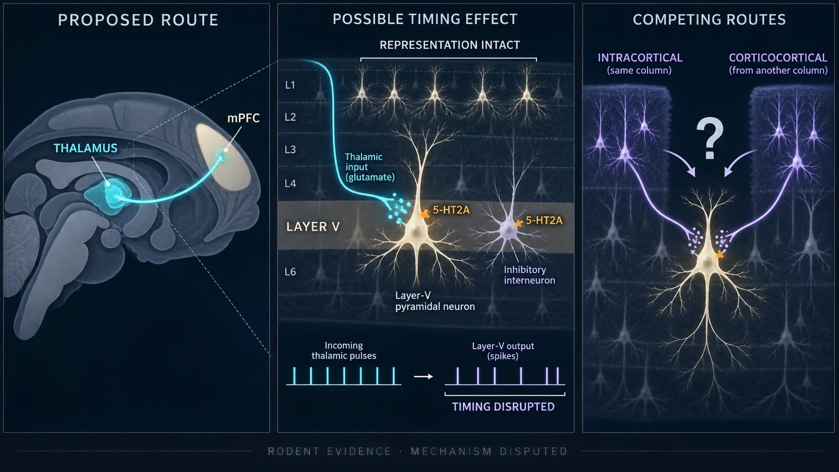
Now the counterweight. A careful study concluded that its results "do not support the idea" that 5-HT2A receptors act on thalamocortical afferents, favoring intracortical networks instead (Béïque, 2007). Others point to corticocortical rather than thalamocortical origins (Liu and Aghajanian, 2008), and to deep rather than superficial layer V (Andrade, 2011).
We have also dropped 2 specifics that earlier drafts of this argument carried: the claim that layer Va in particular gates thalamic input to anterior cortex. Both trace back to a mouse expression paper that was not a thalamocortical study at all. The evidence supports a rodent, prefrontal, superficial layer V statement, and nothing more precise than that.
And one more thing we will not claim
It is often said that 5-HT2A agonism increases gamma oscillations, which would tie neatly to timing. The data do not cooperate. In humans, psilocybin reduced broadband power with no effect on induced gamma (Muthukumaraswamy, 2013); in rat, gamma decreased (Wood, 2012); a later study found only a nonsignificant upward trend (Golden and Chadderton, 2022). The direction is inconsistent, so we make no gamma claim.
Where the receptor story runs out.
The cerebellum is the brain's forward model. It predicts the sensory consequence of a movement, compares the prediction against what happened, and corrects the difference (Wolpert, Miall and Kawato, 1998; Ito, 2008). It is also exquisitely timing dependent (Ivry and Keele, 1989).
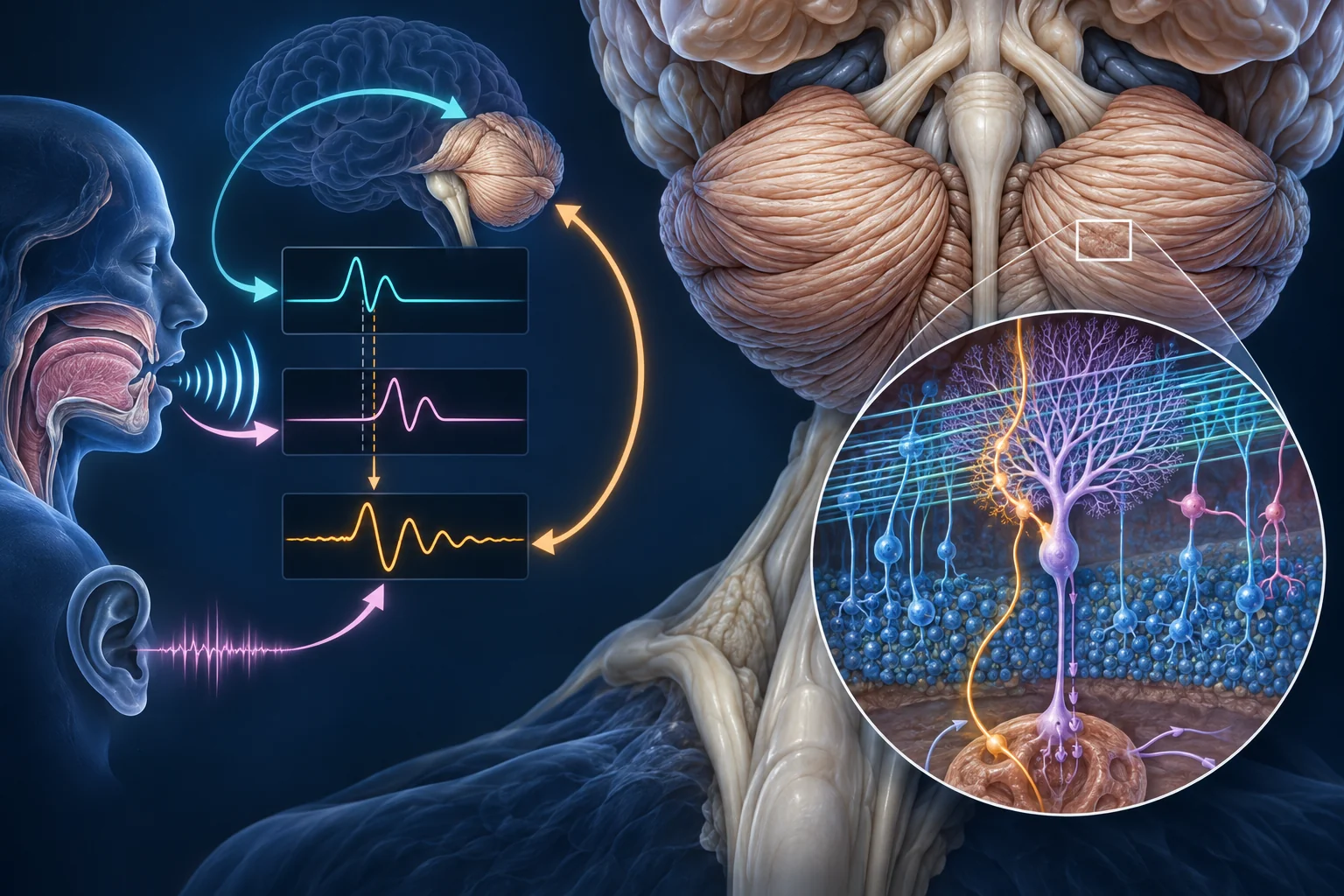
A system built on comparing prediction to outcome is a system that depends on a clean error signal. Feed it a signal that is subtly and endlessly wrong and it cannot calibrate. Damage the cerebellum and adaptation to a perturbed movement fails (Tseng, 2007), with increased motor variability as one contributor (Therrien, 2016), though not the only one (Vassiliadis, 2019).
So the functional logic is clean. The receptor logic is not, and this is where we part company with our own earlier draft.
Bar lengths encode the descriptive categories used by the source authors, not measured values. Cerebellum is reported among regions of only low to very low density (Pazos, 1987), and is described as virtually devoid of 5-HT2A receptors, which is precisely why it is used as the reference region in receptor imaging (Ganz, 2017). Some specific binding has been detected in monkey cerebellum (Watabe, 2000), so "very low" is accurate and "absent" is not.
Which leaves a cleaner competing explanation, and we should say it plainly: the cerebellum may be failing not because it is exposed to more of anything, but because it is less tolerant of noise than cortex is. Timing critical systems degrade first when their inputs get noisy, whatever the source of the noise. That explanation requires no tryptamine at all.
The question worth funding is therefore not "is the cerebellum affected" but differential exposure or differential sensitivity. A dose response relationship would separate them. Nobody has run it. We have also dropped the earlier claim that Purkinje cells carry 5-HT2A receptors, because we could not verify a primary source for it.
What the scanner actually saw.
The strongest body of work on motor circuits in autism comes from Stewart Mostofsky's group at Kennedy Krieger. Each finding below is paired with the authors' own interpretation, which is not always the interpretation this page would prefer.
Mostofsky, "Decreased connectivity and cerebellar activity in autism during motor task performance."
Authors' reading: the reduced cerebellar activation may reflect difficulty shifting execution from cortical regions handling effortful control to regions handling habitual, automatic execution. Note the sample size.
Lidstone, Rochowiak, Mostofsky and Nebel, data driven resting state analysis.
The part usually left out: the same region showed stronger connectivity with primary motor cortex. This is a reorganization, not simply less of something.
Nebel, on functional segregation of motor cortex.
Authors' reading: this "may be due, at least in part" to a delay in functional specialization. The data are cross sectional, so delayed maturation is an inference, not an observation.
McLaughlin, Crocetti, Mostofsky and Lidstone.
Precision matters: left hemisphere only (p = 0.0087; right hemisphere p = 0.33). It tracked praxis performance, not core autism symptom scores.
A related finding on interhemispheric connectivity between right and left somatomotor cortex (Anderson, 2011) is often attributed to this group. It is not theirs: that study came out of the University of Utah. Attribution is part of accuracy.
The methodological problem, raised by the same group
In a later paper from Mostofsky's own lab, 28.5% of autistic children were excluded for head motion versus 16.1% of typically developing children (Nebel, 2022). The children who survive that filter are older, more able and better coordinated than the children who do not. Every imaging result above is drawn from the children who could hold still, which is a systematic bias toward the mildest end of the population this page is about.
And the imaging literature disagrees with itself: increased cerebellar activation has been reported (Allen, 2004), as has hyperconnectivity (Supekar, 2013), and one careful analysis found no whole brain difference at all (Tyszka, 2014).
Why this shows up as behavior.
Motor planning is not a niche skill. It is the final common path for almost everything we use to judge a child.
Speech is the most punishing timing task the human body performs: dozens of muscles sequenced within milliseconds, with no tolerance for drift. Pointing, gesturing, imitating, and following an instruction on request all run through the same planning and release machinery.

Which means a child with intact comprehension and disrupted output will fail nearly every standard measure of comprehension, because every one of those measures is scored on output. The assessment cannot distinguish "does not know" from "cannot show."
It also reframes behavior. A demand placed on a system that cannot reliably release a motor plan produces distress, escape, and what gets recorded as noncompliance or aggression. Read as a planning failure rather than a willingness failure, the behavior becomes predictable.
On speech specifically we have to correct a claim we have made before. Childhood apraxia of speech is not established as common in autism: one study found no statistical support for it in 46 verbal autistic children (Shriberg, 2011), while a clinic sample reported much higher co-occurrence (Tierney, 2015). In minimally verbal children, suspected cases were found in 13 of 54 (Chenausky, 2019). The true rate is unsettled, and no reliability study exists for diagnosing it in minimally verbal children at all.
What about children who spell?
The argument that representation is intact while output is disrupted is often illustrated with children who communicate by spelling on a letterboard. We are not going to use that evidence the way it is usually used, and families deserve to know why.
Facilitated communication, where a facilitator supports a person's hand, has been tested directly and failed. In controlled message passing tests, users typed what the facilitator had been shown rather than what they themselves had seen (Montee, 1995), a pattern confirmed repeatedly (Saloviita, 2014) with reviewers finding unequivocal evidence of facilitator control (Schlosser, 2014). Professional bodies classify it as discredited.
Newer methods, including spelling to communicate and the rapid prompting method, are distinct from facilitated communication, but the evidence for independent authorship is not there yet. 2 systematic reviews looking for qualifying studies of authorship found none (Schlosser, 2019; Schlosser, 2026).
The main evidence offered for agency is indirect: eye tracking in 9 users showed gaze patterns interpreted as consistent with authorship (Jaswal, 2020). That study included no message passing test and the assistant held the board, and it has been criticized on those grounds (Beals, 2021). A later study demonstrated spelling knowledge without assistance (Jaswal, 2024), which tests literacy rather than authorship.
One term should be retired regardless of where the evidence lands: describing any letterboard method as "motor free" is incorrect. Pointing is a motor act. A method built on pointing cannot, by construction, bypass a motor planning problem.
Why the argument survives anyway
The dissociation between intact representation and disrupted output does not depend on letterboard evidence, and should never have been rested on it. It is carried by measurements that require no facilitator at all: praxis deficits that persist after controlling for basic motor skill, the specificity of those deficits compared with ADHD, and imaging of motor circuits in children who were not communicating at the time. That is the evidence this page is built on.
Every link, with its evidence grade.
This is the Endogenous Psychedelic Hypothesis stated as a chain. A chain is only as strong as its weakest link, so here is every link with an honest grade attached.
Gut bacteria decarboxylate tryptophan to tryptamine
EstablishedConfirmed in specific strains of R. gnavus, C. sporogenes, B. hansenii and others. Strain level, not genus level.
Barrier permeability lets it reach the circulation
ContestedPositive in several studies, negative in the largest. Best read as a subgroup finding, not a feature of autism as a group.
Circulating tryptamine is elevated in these children
Never measuredNo published study has measured it in autism. This is the cheapest missing experiment in the chain.
Low activity MAO-A in hemizygous males clears it poorly
Genetics solid, inference untestedThe X linkage and the promoter variants are established. That they produce a measurable tryptamine load in autistic boys is not.
Methylation converts tryptamine to DMT
Enzyme unidentifiedKnockout work shows methylation proceeds without INMT and yields neither N-methyltryptamine nor DMT. The step is real; the enzyme is not identified.
It reaches brain concentrations that matter
UnprovenEndogenous DMT has never been quantified in human brain tissue, and the pharmacology literature holds that trace levels are insufficient.
5-HT2A agonism disturbs gating of motor plans
Rodent, prefrontal, disputedSupported for thalamocortical modulation in rodent prefrontal cortex, and directly contradicted by at least one careful study. No human evidence.
3 links are open and one is contested. That is the accurate state of this model in September 2026. Anyone presenting it as settled science, including us in earlier drafts, is overstating it.
What would prove this wrong.
A hypothesis that cannot be killed is not a hypothesis. Here is what would kill this one, and what would raise it.
| Experiment | Result that would falsify | Result that would support |
|---|---|---|
| Measure circulating tryptamine in autistic and comparison children | No difference in a well powered sample | Elevation that tracks decarboxylase carriage |
| Quantify decarboxylase gene capacity in stool, not genus abundance | Tryptamine output unrelated to total enzyme capacity | Output tracks capacity across taxa, independent of R. gnavus |
| Identify the enzyme that methylates tryptamine in mammals | No mammalian enzyme produces DMT at physiological rates | A named enzyme, with expression in relevant tissue |
| Quantify DMT in human brain tissue | Levels far below receptor relevant concentrations | Levels in the range that occupies 5-HT2A |
| Test 5-HT2A gating of thalamic input in human cortex | Effect is intracortical, not thalamocortical | Thalamocortical timing effect confirmed in human tissue |
| Dose response of tryptamine load against cerebellar and praxis measures | Motor findings unrelated to tryptamine load | Graded relationship, separating exposure from sensitivity |
| Repeat the motor imaging with head motion handled without exclusion | Findings vanish once severely affected children are included | Findings hold, or strengthen, in the children who matter most |
Not one of these is exotic. Several could be done with samples that already sit in freezers. The reason to take this model seriously is not that it is proven. It is that every one of its open links is measurable, which is more than can be said for most theories of autism offered in the last 50 years.
Read the silence correctly.
Strip out every unproven link and something still stands, and it does not depend on any molecule.
Praxis in autism is impaired beyond what coordination explains, and the more impaired it is, the more severe the presentation. Motor circuits in these children are measurably reorganized, with the visual system routing away from sensorimotor cortex and toward prefrontal cortex, and that rerouting tracks praxis rather than symptom scores. Those are not findings about a missing mind. They are findings about a system building a slower, more effortful, more conscious path to the same output, because the automatic one is not reliable.
That is what "he is not responding" looks like from the inside of the network.
That error is not theoretical. It sets the expectations, the placement, the curriculum, and the way a person is spoken to in their own presence for decades. If the motor account is even partly right, a large number of people have been assessed on the one channel they cannot use.
The tryptamine model may turn out to be wrong. 3 of its links are open, and we have marked every one. But the conclusion that matters this week does not wait on any of that: the absence of an answer is not evidence of an absent mind. Presume competence, address the child as the age they are, and make the assessment fit the child rather than the child fit the assessment.
The apraxia was never silence. It was interference on the way out.
The mechanism, on real anatomy.
The argument of this page in 4 minutes, step by step: the gut bacteria that make tryptamine, the receptor in layer 5 of frontal cortex, how the cerebellum corrects a movement, and why it withdraws when the signal stays wrong.
What is real in this film
The brain, the fiber tracts, the neurons and the proteins are real data, not drawings: a standard human brain surface and template, population tractography, reconstructed human neurons, and the published structures of the bacterial enzyme that makes tryptamine, of MAO‑A, and of the 5‑HT2A receptor holding DMT. Every study named on screen is in the reference list below. The vocal tract, the muscle timing chart, the cranial nerve courses and the prediction and error traces are schematic and are labeled as such. 2 things need stating plainly. The line about urinary tryptamines running high is a clinical observation from this practice, not a published or controlled finding, and no study has yet measured circulating tryptamine in autism. And the film draws a contrast with spelling on a letterboard, which the section above deliberately does not rely on, because independent authorship in those methods is not established.
Credits: brain surface, FreeSurfer fsaverage. Template, ICBM152 2009a, Copyright (C) 1993 to 2004 Louis Collins, McConnell Brain Imaging Centre, Montreal Neurological Institute, McGill University. Tractography, Advanced Atlas of 80 Bundles (c) Eleftherios Garyfallidis, CC BY 4.0 (Garyfallidis 2017; Yeh 2018), via DIPY. Neurons, NeuroMorpho.Org, CC BY 4.0 (Tecuatl, Ljungquist and Ascoli 2024), from the Allen Cell Types, Allman, Markram and D’Angelo archives. Structures, RCSB PDB 4OBV, 2Z5X, 9AS2. Atlases, Destrieux, Harvard‑Oxford, AAL.
About this page
This article is educational and is not medical advice, a diagnosis, or a treatment plan. Nothing here should be used to start, stop or change any intervention for a specific child. The Endogenous Psychedelic Hypothesis is a scientific model under active investigation. It is not established fact, and the individual links in it are graded above precisely so that readers can see where the evidence is strong and where it is absent.
The cohort figures quoted are from sequencing data generated in this practice and have not been peer reviewed or published. They are offered as an observation that motivates a testable question, not as evidence of a mechanism. For questions about this article, please contact us.
Related reading on this site: DMT literature summarized, the DMT in Autism study page, and the DMT Research Library, which holds 225 studies in searchable form.
Primary literature cited on this page
- Ackermann H, Mathiak K, Ivry RB. Temporal organization of "internal speech" as a basis for cerebellar modulation of cognitive functions. Behav Cogn Neurosci Rev. 2004;3(1):14. doi:10.1177/1534582304263251
- Allen G, et al. Cerebellar function in autism. Biol Psychiatry. 2004. doi:10.1016/j.biopsych.2004.06.005
- Anderson JS, et al. Decreased interhemispheric functional connectivity in autism. Cereb Cortex. 2011;21(5):1134. doi:10.1093/cercor/bhq190
- Andrade R. Serotonergic regulation of neuronal excitability. Neuropharmacology. 2011. doi:10.1016/j.neuropharm.2011.01.015
- Axelrod J. Enzymatic formation of psychotomimetic metabolites. Science. 1961;134(3475):343. doi:10.1126/science.134.3475.343
- Barker SA. N,N-dimethyltryptamine, a review. Front Neurosci. 2018;12:536. doi:10.3389/fnins.2018.00536
- Barre A, et al. Presynaptic serotonin 2A receptors modulate thalamocortical plasticity. PNAS. 2016;113(10). doi:10.1073/pnas.1525586113
- Beals K. Commentary on eye tracking and assisted communication. Evidence-Based Communication Assessment and Intervention. 2021. doi:10.1080/17489539.2021.1918890
- Béïque JC, et al. Mechanism of the 5-HT2A receptor mediated facilitation of synaptic activity. PNAS. 2007;104(23):9870. doi:10.1073/pnas.0700436104
- Brunner HG, et al. Abnormal behavior associated with a point mutation in monoamine oxidase A. Science. 1993;262:578. doi:10.1126/science.8211186
- Chenausky K, et al. Motor speech impairment in minimally verbal children. Autism & Developmental Language Impairments. 2019. doi:10.1177/2396941519856333
- Dalton N, et al. Gut permeability in autism spectrum disorders. Autism Res. 2013;7(3):305. doi:10.1002/aur.1350
- de Magistris L, et al. Alterations of the intestinal barrier. JPGN. 2010. doi:10.1097/MPG.0b013e3181dcc4a5
- Dean JG, et al. Biosynthesis and extracellular concentrations of DMT in mammalian brain. Sci Rep. 2019;9. doi:10.1038/s41598-019-45812-w
- D'Eufemia P, et al. Abnormal intestinal permeability in autism. Acta Paediatr. 1996. doi:10.1111/j.1651-2227.1996.tb14220.x
- Dziuk MA, et al. Dyspraxia in autism. Dev Med Child Neurol. 2007;49(10):734. doi:10.1111/j.1469-8749.2007.00734.x
- Esnafoglu E, et al. Increased serum zonulin levels in children with autism. J Pediatr. 2017. doi:10.1016/j.jpeds.2017.04.004
- Fiorentino M, et al. Blood brain and intestinal epithelial barrier alterations in autism. Mol Autism. 2016;7. doi:10.1186/s13229-016-0110-z
- Fournier KA, et al. Motor coordination in autism, a meta-analysis. J Autism Dev Disord. 2010. doi:10.1007/s10803-010-0981-3
- Ganz M, et al. Cerebellar 5-HT2A receptor binding. J Cereb Blood Flow Metab. 2017;37(9):3243. doi:10.1177/0271678x16686092
- Gevi F, et al. Urinary metabolomics of young Italian autistic children. Mol Autism. 2016;7. doi:10.1186/s13229-016-0109-5
- Glynos NG, et al. INMT knockout and endogenous tryptamine methylation. Sci Rep. 2023;13. doi:10.1038/s41598-023-27538-y
- Golden CE, Chadderton P. Psilocybin and cortical oscillations. Sci Rep. 2022;12. doi:10.1038/s41598-022-16325-w
- Gonçalves S, et al. Bacterial tryptophan metabolism. Front Microbiol. 2022;13. doi:10.3389/fmicb.2022.873555
- Gumpper RH, et al. The structural diversity of psychedelic drug actions revealed. Nat Commun. 2025;16:2734. doi:10.1038/s41467-025-57956-7
- Ito M. Cerebellar long term depression: characterization, signal transduction, and functional roles. Physiol Rev. 2001;81(3):1143. doi:10.1152/physrev.2001.81.3.1143
- Ito M. Control of mental activities by internal models in the cerebellum. Nat Rev Neurosci. 2008. doi:10.1038/nrn2332
- Ivry RB, Keele SW. Timing functions of the cerebellum. J Cogn Neurosci. 1989;1(2):136. doi:10.1162/jocn.1989.1.2.136
- Jakab RL, Goldman-Rakic PS. 5-HT2A serotonin receptor in the primate cerebral cortex. J Comp Neurol. 2000;417(3):337. PMID 10683608
- Jaswal VK, Wayne A, Golino H. Eye tracking reveals agency in assisted autistic communication. Sci Rep. 2020;10. doi:10.1038/s41598-020-64553-9
- Kot M, et al. Monoamine oxidase isoforms and trace amine metabolism. Curr Neuropharmacol. 2025;23(11). doi:10.2174/011570159X340635241022113450
- Lambe EK, Aghajanian GK. Serotonin and thalamocortical inputs. J Neurosci. 2001;21(24):9955.
- Levy ER, et al. Localization of human monoamine oxidase A gene. Genomics. 1989. doi:10.1016/0888-7543(89)90072-4
- Lidstone DE, Rochowiak R, Mostofsky SH, Nebel MB. A data driven approach to cerebellar connectivity in autism. Autism Res. 2021. doi:10.1002/aur.2476
- Liu RJ, Aghajanian GK. Corticocortical origin of serotonin induced excitatory postsynaptic currents. PNAS. 2008. doi:10.1073/pnas.0706679105
- MacNeil LK, Mostofsky SH. Specificity of dyspraxia in autism. Neuropsychology. 2012. doi:10.1037/a0026955
- McLaughlin K, Crocetti D, Mostofsky SH, Lidstone DE. Visual to motor connectivity and praxis in autism. Autism Res. 2026;19(2). doi:10.1002/aur.70146
- Montee BB, et al. An experimental analysis of facilitated communication. J Appl Behav Anal. 1995;28(2):189. doi:10.1901/jaba.1995.28-189
- Mostofsky SH, et al. Decreased connectivity and cerebellar activity in autism during motor task performance. Brain. 2009;132(9):2413. doi:10.1093/brain/awp088
- Muthukumaraswamy SD, et al. Broadband cortical desynchronization underlies psilocybin effects. J Neurosci. 2013;33(38):15171. doi:10.1523/jneurosci.2063-13.2013
- Nebel MB, et al. Precentral gyrus functional connectivity signatures of autism. Hum Brain Mapp. 2014;35(2):567. doi:10.1002/hbm.22188
- Nebel MB, et al. Accounting for motion in resting state fMRI. NeuroImage. 2022;257:119296.
- Nichols DE. N,N-dimethyltryptamine and the pineal gland. J Psychopharmacol. 2017;32(1):30. doi:10.1177/0269881117736919
- Otaru N, et al. Intestinal bacteria derived tryptamine. Front Microbiomes. 2024;3. doi:10.3389/frmbi.2024.1373335
- Ozelius L, et al. Human monoamine oxidase gene localization. Genomics. 1988. doi:10.1016/0888-7543(88)90159-0
- Pazos A, Probst A, Palacios JM. Serotonin receptors in the human brain. Neuroscience. 1987;21(1):123. doi:10.1016/0306-4522(87)90327-7
- Robertson MA, et al. Intestinal permeability and autism. J Autism Dev Disord. 2008. doi:10.1007/s10803-007-0482-1
- Sabol SZ, Hu S, Hamer D. A functional polymorphism in the monoamine oxidase A promoter. Hum Genet. 1998;103(3):273. doi:10.1007/s004390050816
- Saloviita T, et al. Authorship in facilitated communication. AAC. 2014;30(3):213.
- Santana N, et al. Expression of 5-HT1A and 5-HT2A receptors in pyramidal and GABAergic neurons. Cereb Cortex. 2004;14(10):1100. doi:10.1093/cercor/bhh070
- Scheffler L, et al. Widely used commercial ELISA does not detect precursor haptoglobin 2. Front Endocrinol. 2018;9:22. doi:10.3389/fendo.2018.00022
- Schlosser RW, et al. Facilitated communication and authorship, a systematic review. AAC. 2014;30(4):359.
- Schlosser RW, et al. Rapid prompting method, a systematic review. Rev J Autism Dev Disord. 2019. doi:10.1007/s40489-019-00175-w
- Shriberg LD, et al. Childhood apraxia of speech in autism. J Autism Dev Disord. 2011;41(4):405. doi:10.1007/s10803-010-1117-5
- Simonyan K, Horwitz B. Laryngeal motor cortex and control of speech in humans. Neuroscientist. 2011;17(2):197. doi:10.1177/1073858410386727
- Son SY, et al. Structure of human monoamine oxidase A at 2.2 A resolution: the control of opening the entry for substrates and inhibitors. Proc Natl Acad Sci U S A. 2008;105(15):5739. doi:10.1073/pnas.0710626105
- Supekar K, et al. Brain hyperconnectivity in children with autism. Cell Rep. 2013. doi:10.1016/j.celrep.2013.10.001
- Therrien AS, Wolpert DM, Bastian AJ. Effective reinforcement learning following cerebellar damage. Brain. 2016;139(1):101. doi:10.1093/brain/awv329
- Thompson MA, et al. Human indolethylamine N-methyltransferase, cloning. J Biol Chem. 1998;273(51):34502. doi:10.1074/jbc.273.51.34502
- Thompson MA, et al. Human indolethylamine N-methyltransferase, tissue expression. Genomics. 1999. doi:10.1006/geno.1999.5960
- Tierney C, et al. Motor speech disorders in autism. J Dev Behav Pediatr. 2015. doi:10.1097/DBP.0000000000000189
- Tseng YW, et al. Sensory prediction errors drive cerebellum dependent adaptation. J Neurophysiol. 2007;98(1):54. doi:10.1152/jn.00266.2007
- Tsugeno Y, Ito A. A key amino acid responsible for substrate selectivity of monoamine oxidase. J Biol Chem. 1997;272(22):14033. doi:10.1074/jbc.272.22.14033
- Tyszka JM, et al. Largely typical patterns of resting state functional connectivity in autism. Cereb Cortex. 2014. doi:10.1093/cercor/bht040
- Vassiliadis P, et al. Reward boosts reinforcement based motor learning. eNeuro. 2019;6(1).
- Watabe H, et al. Quantitative analysis of 5-HT2A receptor binding. J Cereb Blood Flow Metab. 2000;20(6):899. doi:10.1097/00004647-200006000-00002
- Williams BB, et al. Discovery and characterization of gut microbiota decarboxylases that produce tryptamine. Cell Host Microbe. 2014;16(4):495. doi:10.1016/j.chom.2014.09.001
- Wolpert DM, Miall RC, Kawato M. Internal models in the cerebellum. Trends Cogn Sci. 1998;2(9):338. doi:10.1016/s1364-6613(98)01221-2
- Wood J, et al. Psychedelic drug effects on cortical oscillations. J Neurosci. 2012;32(9):3022. doi:10.1523/jneurosci.6377-11.2012
- Yu AM, et al. Human cytochrome P450 and monoamine oxidase mediated tryptamine metabolism. J Pharmacol Exp Ther. 2003;304(2). doi:10.1124/jpet.102.043786
- Zhang C, Marek GJ. Thalamic afferents and 5-HT2A receptor mediated responses. J Pharmacol Exp Ther. 2007;320(1):437. doi:10.1124/jpet.106.107490